Breast Cancer Lymph Node Color Doppler Ultrasound Color Meanings

Normal Lymph Node On Ultrasound Lymph Nodes Typically Are Smooth Download Scientific Diagram

Normal Lymph Node On Ultrasound Lymph Nodes Typically Are Smooth Download Scientific Diagram

Color Doppler Sonography Characterizing Breast Lesions

Color Doppler Sonography Characterizing Breast Lesions

Typical Us Appearance Of Lymph Nodes Typical Reactive Node Image A Download Scientific Diagram

Typical Us Appearance Of Lymph Nodes Typical Reactive Node Image A Download Scientific Diagram

Reactive Vs Malignant Lymph Nodes Ultrasound Features Radiology Reference Article Radiopaedia Org

Reactive Vs Malignant Lymph Nodes Ultrasound Features Radiology Reference Article Radiopaedia Org

Pdf Ultrasonography Of Superficial Lymph Nodes Benign Vs Malignant

Pdf Ultrasonography Of Superficial Lymph Nodes Benign Vs Malignant

Ultrasound Evaluation Of Regional Lymph Nodes As An Extension Of The Breast Ultrasound Exam Youtube

Ultrasound Evaluation Of Regional Lymph Nodes As An Extension Of The Breast Ultrasound Exam Youtube

Ultrasound Evaluation Of Regional Lymph Nodes As An Extension Of The Breast Ultrasound Exam Youtube

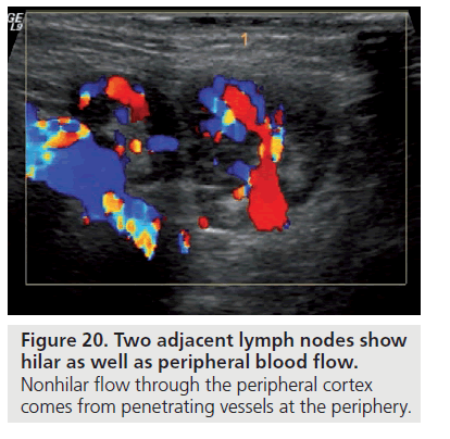
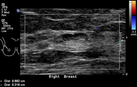
On color doppler it may show some peripheral vascularity in acute phase and may resemble breast cancer requiring a biopsy to establish tissue diagnosis.

Breast cancer lymph node color doppler ultrasound color meanings.

Thus for some time aln dissection alnd was the standard of care in these patients however screening programs and increased awareness on the disease tend to identify more breast cancer patients at an earlier stage in. Several gray scale and color doppler features favor malignancy in a lymph node. As lymph fluid leaves the breast and eventually goes back into the bloodstream the lymph nodes try to catch and trap cancer cells before they reach other parts of the body. Lymphadenopathyis quite common and it can be very difficult to differentiate malignant lymphadenopathy from reactive nodal enlargement.

The longitudinal transverse axis ratio and hilar status on color doppler flow and gray scale us images were prospectively studied for each of 145 axillary nodes in 135 women 74. Color doppler ultrasound can show flow in all lymph nodes regardless of whether they are affected by a benign or a malignant process. The vascular flow is inadequately evaluated. Gray scale parameters that favor malignancy size.

However fat necrosis usually exhibits no significant blood flow figure 16. Axillary lymph node dissection alnd is the definitive method to diagnose axillary metastasis but sentinel lymph node biopsy slnb has supplanted this procedure as the primary method of evaluating the axilla in most cases of early stage breast cancer because slnb has a significantly lower rate of morbidity than does alnd and a low false. Meanwhile research conducted by yang et al. In 1998 reports that the use of color doppler sonography on axillary lymph nodes to diagnose the metastatic axillary lymph nodes of breast cancer.

Lymph nodes are small bean shaped organs that act as filters along the lymph fluid channels. The analysis of patterns of nodal vascularity can be used to differentiate benign from malignant lymphadenopathy with high sensitivity. Axillary lymph node aln status is the most important prognostic factor for the management of breast cancer patients in the absence of metastatic disease. B d three dimensional multiplanar reformatted images also show loss of the fatty hilum.

The presence of cancer cells is known as lymph node involvement.

Breast Cancer Topic Lymph Nodes On Ultrasound

Breast Cancer Topic Lymph Nodes On Ultrasound

Intramammary Lymph Nodes Radiology Reference Article Radiopaedia Org

Intramammary Lymph Nodes Radiology Reference Article Radiopaedia Org

Lymph Node Matting Multiple Malignant Nodes Are Fused In A Single Ill Download Scientific Diagram

Lymph Node Matting Multiple Malignant Nodes Are Fused In A Single Ill Download Scientific Diagram

Nodal Echogenic Hilum A Benign Node With Echogenic Central Hilum B Download Scientific Diagram

Nodal Echogenic Hilum A Benign Node With Echogenic Central Hilum B Download Scientific Diagram

Source : pinterest.com